


PRODUCTS
With proprietary intellectual property of MicroStacker™ and SuperISH™,
we have a market-leading portfolio of IHC and ISH reagents and instruments.
-
 I
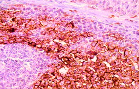
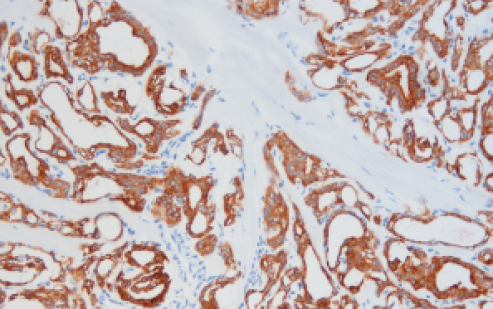
IIMMUNOHISTOCHEMISTRY
-
 I
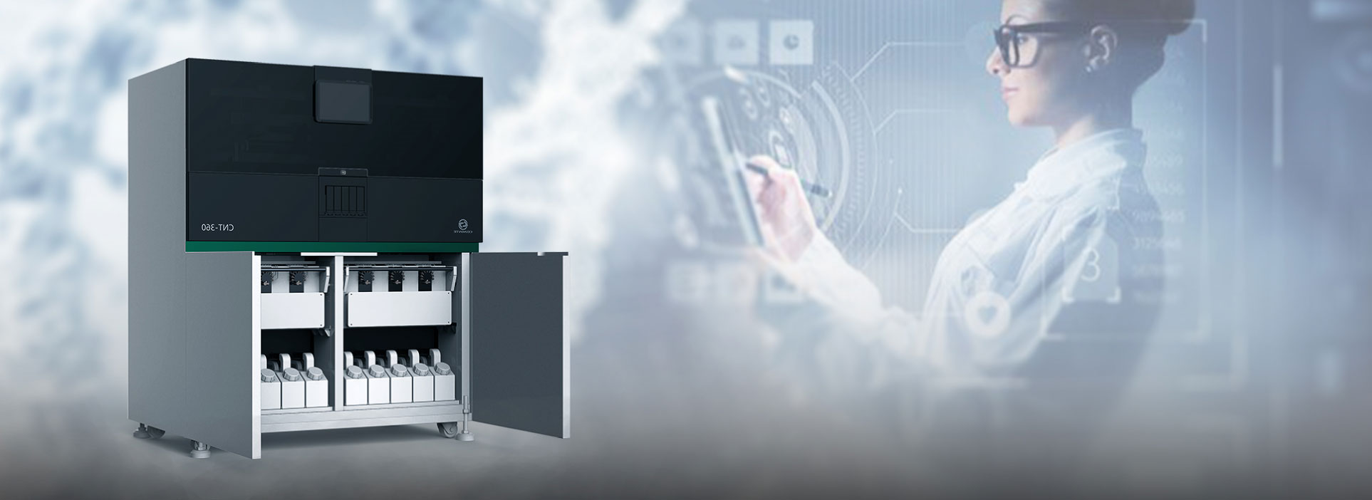
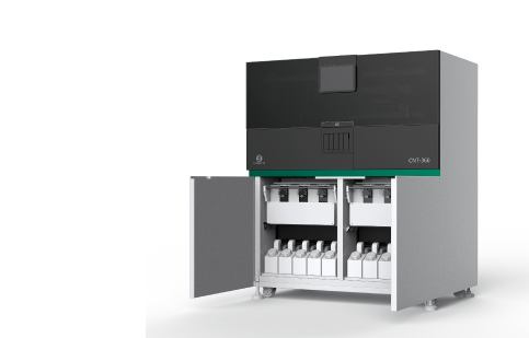
IINSTRUMENT
-
 I
IMOLECULAR
-
 I
IFROZEN SECTION IHC
-
 I
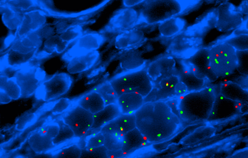
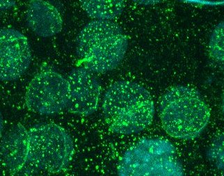
IIMMUNOFLUPRESCENCE
-
 I
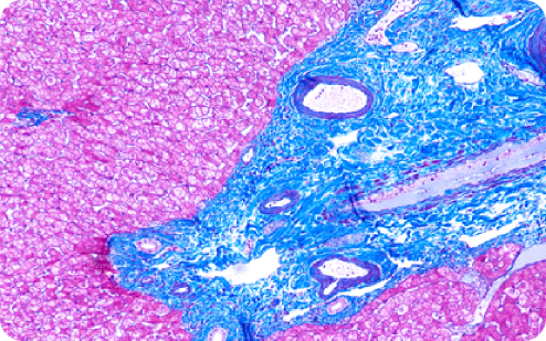
IHISTOLOGY
-
 I
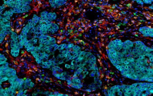
IIMMUNOCYTOCHEMISTRY
-
 I
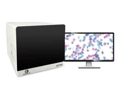
IDIGITAL PATHOLOGY
SOLUTIONS
We provide Clinical Solutions, R&D Collaboration and OEM/ODM Partnership.


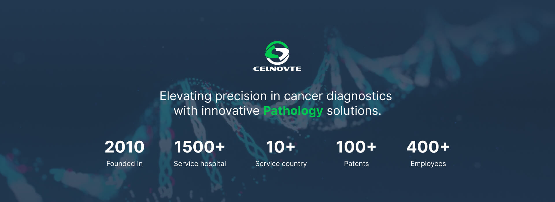
FACILITIES & CERTIFICATES
Our facilities in China are NMPA & GMP compliant
and certified for ISO13485 and ISO9001.


 on Automatic IHC stainer CNT360 is honored as recommendation from NordiQC")

 0
0
